martes, 11 de diciembre de 2007


Circulatory system:


The circulatory system (or cardiovascular system) is an organ system that moves nutrients, gases, and wastes to and from cells, and helps stabilize body temperature and pH to maintain homeostasis. While humans, as well as other vertebrates have a closed circulatory system, some invertebrate groups have open circulatory system. The most primitive animal phyla lack circulatory systems.

Human circulatory system:
The main components of the human circulatory system are the heart, the blood, and the blood vessels.
Furthermore, these components can either belong to the systemic circulation and the pulmonary circulation. The systemic circulation is the main part of the circulatory system, while the pulmonary system oxygenates the blood.

Systemic circulation:
Systemic circulation is the portion of the cardiovascular system which carries oxygenated blood away from the heart, to the body, and returns deoxygenated blood back to the heart.
Arteries always take blood away from the heart, regardless of their oxygenation, and veins always bring blood back. In general, arteries bring oxygenated blood to the tissues; veins bring deoxygenated blood back to the heart. In the case of the pulmonary vessels, however, the oxygenation is reversed: the pulmonary artery takes deoxygenated blood from the heart to the lungs, and oxygenated blood is pumped back through the pulmonary vein to the heart. As blood circulates through the body, oxygen and nutrients diffuse from the blood into cells surrounding the capillaries, and carbon dioxide diffuses into the blood from the capillary cells.
The release of oxygen from red blood cells or erythrocytes is regulated in mammals. It increases with an increase of carbon dioxide in tissues, an increase in temperature, or a decrease in pH. Such characteristics are exhibited by tissues undergoing high metabolism, as they require increased levels of oxygen.

Pulmonary circulation:
Main article: Pulmonary circulation
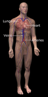
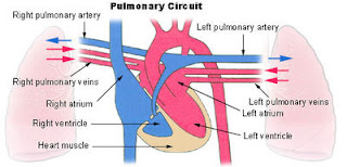
Pulmonary circulation is the portion of the cardiovascular system which carries oxygen-depleted blood away from the heart, to the lungs, and returns oxygenated blood back to the heart.
De-oxygenated blood enters the right atrium of the heart and flows into the right ventricle where it is pumped through the pulmonary arteries to the lungs. Pulmonary veins return the now oxygen-rich blood to the heart, where it enters the left atrium before flowing into the left ventricle. From the left ventricle the oxygen-rich blood is pumped out via the aorta, and on to the rest of the body.


Main article: Systemic circulation


Systemic circulation is the portion of the cardiovascular system which carries oxygenated blood away from the heart, to the body, and returns deoxygenated blood back to the heart. The term is contrasted with pulmonary circulation.


Main article: Pulmonary circulation

Pulmonary circulation: is the portion of the cardiovascular system which carries oxygen-depleted blood away from the heart, to the lungs, and returns oxygenated blood back to the heart. The term is contrasted with systemic circulation.


Respiratory system:
Among quadrupeds, the respiratory system generally includes tubes, such as the bronchi, used to carry air to the lungs, where gas exchange takes place. A diaphragm pulls air in and pushes it out. Respiratory systems of various types are found in a wide variety of organisms.
In humans and other mammals, the respiratory system consists of the airways, the lungs, and the respiratory muscles that mediate the movement of air into and out of the body. Within the alveolar system of the lungs, molecules of oxygen and carbon dioxide are passively exchanged, by diffusion, between the gaseous environment and the blood. Thus, the respiratory system facilitates oxygenation of the blood with a concomitant removal of carbon dioxide and other gaseous metabolic wastes from the circulation. The system also helps to maintain the acid-base balance of the body through the efficient removal of carbon dioxide from the blood.

Anatomy:
In humans and other animals, the respiratory system can be conveniently subdivided into an upper respiratory tract (or conducting zone) and lower respiratory tract (respiratory zone), trachea and lungs.
Air moves through the body in the following order:

-Pharynx (naso-, oro-, laryngo-) .
-Larynx (voice box)


Pharynx:


The pharynx (plural: pharynges) is the part of the neck and throat situated immediately posterior to the mouth and nasal cavity, and cranial, or superior, to the esophagus, larynx, and trachea.


miércoles, 21 de noviembre de 2007


Tissue:

Biological tissue is a collection of interconnected cells that perform a similar function within an organism.
The study of tissue is known as histology, or, in connection with disease, histopathology.
The classical tools for studying the tissues are the wax block, the tissue stain, and the optical microscope, though developments in electron microscopy, immunofluorescence, and frozen sections have all added to the sum of knowledge in the last couple of decades.With these tools, the classical appearances of the tissues can be examined in health and disease, enabling considerable refinement of clinical diagnosis and prognosis. There are four basic types of tissue in the body of all animals, including the human body and lower multicellular organisms such as insects. These compose all the organs, structures and other contents.
Epithelium - Tissues composed of layers of cells that cover organ surfaces such as surface of the skin and inner lining of digestive tract: the tissues that serve for protection, secretion, and absorption.
Connective tissue - As the name suggests, connective tissue holds everything together. Connective tissue is characterized by the separation of the cells by an inorganic material, which is called extracellular matrix. Bone and blood are connective tissues.
Muscle tissue - Muscle cells contain contractile filaments that move past each other and change the size of the cell. Muscle tissue also is separated into three distinct categories: visceral or smooth muscle, which is found in the inner linings of organs; skeletal muscle, which is found attached to bone in order for mobility to take place; and cardiac muscle which is found in the heart.
Nervous tissue - Cells forming the brain, spinal cord and peripheral nervous system.

Plant tissues:
Examples of tissue in other multicellular organisms are vascular tissue in plants, such as xylem and phloem. Plant tissues are categorized broadly into three tissue systems: the epidermis, the ground tissue, and the vascular tissue. Together they are often referred to as biomass.
Epidermis - Cells forming the outer surface of the leaves and of the young plant body.
Vascular tissue - The primary components of vascular tissue are the xylem and phloem. These transport fluid and nutrients internally.
Ground tissue - Ground tissue is less differentiated than other tissues. Ground tissue manufactures nutrients by photosynthesis and stores reserve nutrients.


Epithelium:


In biology and medicine, epithelium is a tissue composed of layers of cells which line the cavities and surfaces of structures throughout the body. It is also the type of tissue of which many glands are formed. Epithelium lines both the outside (skin) and the inside cavities and lumen of bodies. The outermost layer of our skin is composed of dead stratified squamous, keratinized epithelial cells.
Mucous membranes lining the inside of the mouth, the oesophagus, and part of the rectum are lined by nonkeratinized stratified squamous epithelium. Other, open to outside body cavities are lined by simple squamous or columnar epithelial cells.
Other epithelial cells line the insides of the lungs, the gastrointestinal tract, the reproductive and urinary tracts, and make up the exocrine and endocrine glands. The outer surface of the cornea is covered with fast-growing, easily-regenerated epithelial cells.
Functions of epithelial cells include secretion, absorption, protection, transcellular transport, sensation detection, and selective permeability.


Connective tissue:


Connective tissue is one of the four types of tissue in traditional classifications (the others being epithelial, muscle, and nervous tissue.) It is largely a category of exclusion rather than one with a precise definition, but all or most tissues in this category are similarly:


Muscle:


Muscle (from Latin musculus, diminutive of mus "mouse"[1]) is contractile tissue of the body and is derived from the mesodermal layer of embryonic germ cells. It is classified as skeletal, cardiac, or smooth muscle[2], and its function is to produce force and cause motion, either locomotion or movement within internal organs. Much of muscle contraction occurs without conscious thought and is necessary for survival, like the contraction of the heart, or peristalsis (which pushes food through the digestive system). Voluntary muscle contraction is used to move the body, and can be finely controlled, like movements of the eye, or gross movements like the quadriceps muscle of the thigh. There are two broad types of voluntary muscle fibers, slow twitch and fast twitch. Slow twitch fibers contract for long periods of time but with little force while fast twitch fibers contract quickly and powerfully but fatigue very rapidly.


Nervous tissue:


-Nervous tissue is the fourth major class of vertebrate tissue. The function of the nervous tissue is in communication between parts of the body. It is composed of neurons, which transmit impulses, and the neuroglia, which assist propagation of the nerve impulse as well as provide nutrients to the neuron.
All nervous tissue of an organism makes up its nervous system, which may include the brain, spinal cord, and nerves throughout the organism.
Nervous tissue is made of nerve cells that come in many varieties, all of which are distinctly characteristic by the axon or long stem like part of the cell that sends action potential signals to the next cell.


2. The Digestive Apparatus;
1.The Digestive Tube (alimentary canal) is a musculomembranous tube, about 9 metres long, extending from the mouth to the anus, and lined throughout its entire extent by mucous membrane. It has received different names in the various parts of its course: at its commencement is the mouth, where provision is made for the mechanical division of the food (mastication), and for its admixture with a fluid secreted by the salivary glands (insalivation); beyond this are the organs of deglutition, the pharynx and the esophagus, which convey the food into the stomach, in which it is stored for a time and in which also the first stages of the digestive process take place; the stomach is followed by the small intestine, which is divided for purposes of description into three parts, the duodenum, the jejunum, and ileum. In the small intestine the process of digestion is completed and the resulting products are absorbed into the blood and lacteal vessels. Finally the small intestine ends in the large intestine, which is made up of cecum, colon, rectum, and anal canal, the last terminating on the surface of the body at the anus.


2.The accessory organs are the teeth, for purposes of mastication; the three pairs of salivary glands—the parotid, submaxillary, and sublingual—the secretion from which mixes with the food in the mouth and converts it into a bolus and acts chemically on one of its constituents; the liver and pancreas, two large glands in the abdomen, the secretions of which, in addition to that of numerous minute glands in the walls of the alimentary canal, assist in the process of digestion.


3.The Development of the Digestive Tube.—The primitive digestive tube consists of two parts, viz.: (1) the fore-gut, within the cephalic flexure, and dorsal to the heart; and (2) the hind-gut, within the caudal flexure (Fig. 977). Between these is the wide opening of the yolk-sac, which is gradually narrowed and reduced to a small foramen leading into the vitelline duct. At first the fore-gut and hind-gut end blindly. The anterior end of the fore-gut is separated from the stomodeum by the buccopharyngeal membrane (Fig. 977); the hind-gut ends in the cloaca, which is closed by the cloacal membrane.


The Mouth.—The mouth is developed partly from the stomodeum, and partly from the floor of the anterior portion of the fore-gut. By the growth of the head end of the embryo, and the formation of the cephalic flexure, the pericardial area and the buccopharyngeal membrane come to lie on the ventral surface of the embryo. With the further expansion of the brain, and the forward bulging of the pericardium, the buccopharyngeal membrane is depressed between these two prominences. This depression constitutes the stomodeum (Fig. 977). It is lined by ectoderm, and is separated from the anterior end of the fore-gut by the buccopharyngeal membrane. This membrane is devoid of mesoderm, being formed by the apposition of the stomodeal ectoderm with the fore-gut entoderm; at the end of the third week it disappears, and thus a communication is established between the mouth and the future pharynx. No trace of the membrane is found in the adult; and the communication just mentioned must not be confused with the permanent isthmus faucium. The lips, teeth, and gums are formed from the walls of the stomodeum, but the tongue is developed in the floor of the pharynx


The Salivary Glands.—The salivary glands arise as buds from the epithelial lining of the mouth; the parotid appears during the fourth week in the angle between the maxillary process and the mandibular arch; the submaxillary appears in the sixth week, and the sublingual during the ninth week in the hollow between the tongue and the mandibular arch.


FIG. 978– Head end of human embryo of about thirty to thirty-one days. (From model by Peters.)


FIG. 979– Floor of pharynx of human embryo about twenty-six days old. (From model by Peters.)

FIG. 980– Floor of pharynx of human embryo of about the end of the fourth week. (From model by Peters.) (See enlarged image)


The Tongue (Figs. 979 to 981).—The tongue is developed in the floor of the pharynx, and consists of an anterior or buccal and a posterior or pharyngeal part which are separated in the adult by the V-shaped sulcus terminalis. During the third week there appears, immediately behind the ventral ends of the two halves of the mandibular arch, a rounded swelling named the tuberculum impar, which was described by His as undergoing enlargement to form the buccal part of the tongue. More recent researches, however, show that this part of the tongue is mainly, if not entirely, developed from a pair of lateral swellings which rise from the inner surface of the mandibular arch and meet in the middle line. The tuberculum impar is said to form the central part of the tongue immediately in front of the foramen cecum, but Hammar insists that it is purely a transitory structure and forms no part of the adult tongue. From the ventral ends of the fourth arch there arises a second and larger elevation, in the center of which is a median groove or furrow. This elevation was named by His the furcula, and is at first separated from the tuberculum impar by a depression, but later by a ridge, the copula, formed by the forward growth and fusion of the ventral ends of the second and third arches. The posterior or pharyngeal part of the tongue is developed from the copula, which extends forward in the form of a V, so as to embrace between its two limbs the buccal part of the tongue. At the apex of the V a pit-like invagination occurs, to form the thyroid gland, and this depression is represented in the adult by the foramen cecum of the tongue. In the adult the union of the anterior and posterior parts of the tongue is marked by the V-shaped sulcus terminalis, the apex of which is at the foramen cecum, while the two limbs run lateralward and forward, parallel to, but a little behind, the vallate papillæ.

FIG. 981– Floor of pharynx of human embryo about thirty days old. (From model by Peter.) (See enlarged image)


The Palatine Tonsils.—The palatine tonsils are developed from the dorsal angles of the second branchial pouches. The entoderm which lines these pouches grows in the form of a number of solid buds into the surrounding mesoderm. These buds become hollowed out by the degeneration and casting off of their central cells, and by this means the tonsillar crypts are formed. Lymphoid cells accumulate around the crypts, and become grouped to form the lymphoid follicles; the latter, however, are not well-defined until after birth.

FIG. 982– Sketches in profile of two stages in the development of the human digestive tube. (His.) A X 30. B X 20. (See enlarged image)